
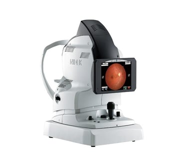
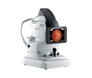
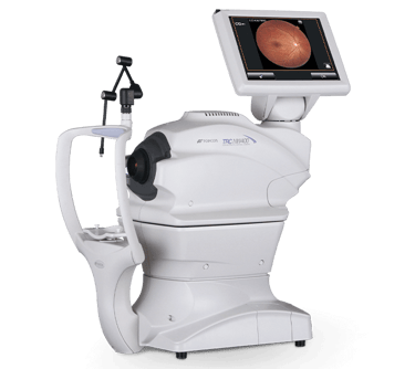
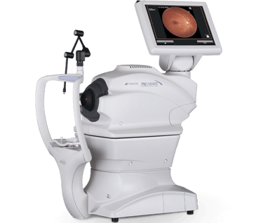
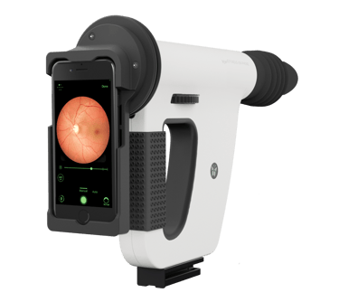
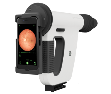
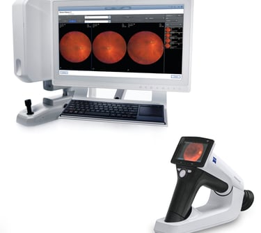
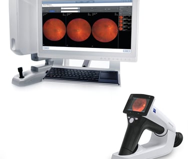
Fundus Camera
A fundus camera is a specialized type of camera used by eye care professionals to capture detailed images of the fundus of the eye. The fundus is the interior surface of the eye, including the retina, optic disc, macula, and blood vessels. These images are crucial for diagnosing and monitoring various eye conditions, such as diabetic retinopathy, glaucoma, macular degeneration, and other retinal diseases.
Here's how it works:
The patient typically sits in front of the camera, and their chin is placed on a support to keep their head still.
The camera takes high-resolution photos of the back of the eye using a special light source.
The images can be used to analyze the health of the retina and the optic nerve, and the doctor can identify issues such as abnormal blood vessels, bleeding, swelling, or signs of disease.
Fundus cameras may also be equipped with various features, such as red-free filters (which enhance the contrast of the blood vessels) or fluorescein angiography, where a dye is injected into the bloodstream to help highlight specific areas of the retina.
Coimbatore, Tamilnadu
Contacts
aeyeswin@hotmail.com